

Lumbar Discectomy for Herniated Disc Relief
This ultra-minimally invasive endoscopic procedure removes fragments of a herniated disc in your spine. This relieves pressure on nearby spinal nerves and can help reduce pain, numbness, and weakness in one or both legs.
What Is a Discectomy?
Discs act as spinal shock absorbers. Of the 23 discs in the spine, 5 are in the lower back. Over time or due to injury, a disc’s outer layer can weaken. This causes the inner material to bulge or herniate, pressing on nerves and causing pain. A discectomy is designed to remove the protruding part to relieve pressure, ease pain, and improve mobility.
Which Conditions Can a Discectomy Address?
Discectomies most commonly address pain related to herniated discs. Common symptoms of a herniated disc include:
- Leg pain that may be accompanied by back pain
- Pain that is felt even when seated
- Numbness, weakness, and/or tingling in one or both legs
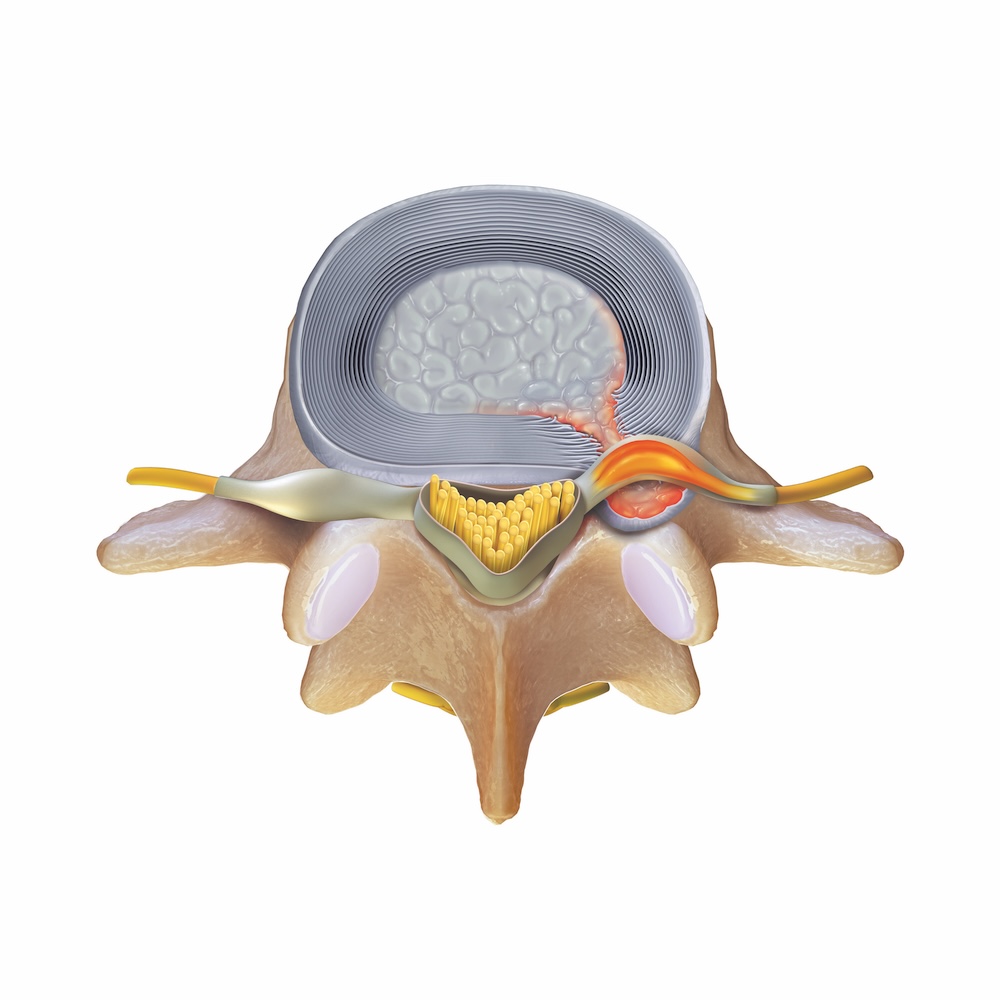
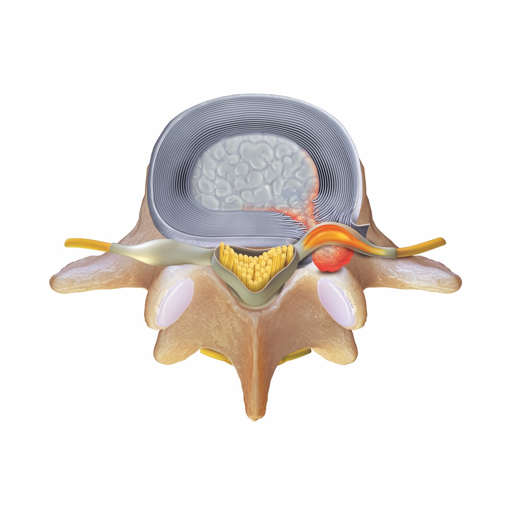

Types of Herniated Discs
Contained Herniation
Extruded Herniation
Sequestered Herniation
Approaches to Endoscopic Lumbar Discectomy
There are two primary approaches to endoscopic lumbar discectomy: interlaminar and transforaminal.
The interlaminar approach is commonly used to address herniations in the lower area of the lumbar spine, generally at the L4-L5 and L5-S1 levels. The transforaminal approach is often used for herniated discs higher in the spine, such as at the L1-L2, L2-L3, and L3-L4 levels. However, your surgeon may recommend this approach at any spinal level based on where the problem is located and your individual anatomy.


Interlaminar Approach
