Lumbar Discectomy for Herniated Disc Relief
This ultra‑minimally invasive endoscopic procedure removes damaged disc fragments to relieve nerve pressure and help reduce pain, numbness, or weakness in one or both legs.
What Is a Discectomy?
Discs act as spinal shock absorbers. Of the 23 discs in the spine, 5 are in the lower back. Over time or due to injury, a disc’s outer layer can weaken. This causes the inner material to bulge or herniate, pressing on nerves and causing pain. A discectomy is designed to remove the protruding part to relieve pressure, ease pain, and improve mobility.
Which Conditions Can a Discectomy Address?
Discectomies most commonly address pain related to herniated discs. Common symptoms of a herniated disc include:
- Leg pain that may be accompanied by back pain
- Pain that is felt even when seated
- Numbness, weakness, and/or tingling in one or both legs
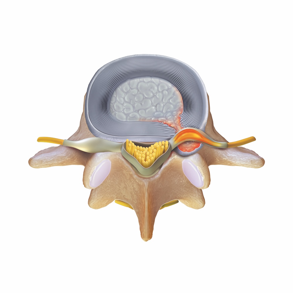
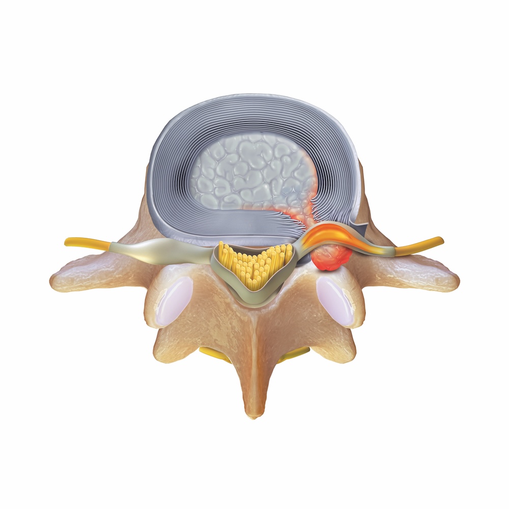

Types of Herniated Discs
Contained Herniation
Nucleus is bulging or stretching the outer annulus but has not pushed through the annulus wall into the spinal canal
Extruded Herniation
Nucleus has broken through the annulus wall, entering the spinal canal, but is still attached to the main body of the disc
Approaches to Endoscopic Lumbar Discectomy
There are two primary approaches to endoscopic lumbar discectomy: interlaminar and transforaminal.
The interlaminar approach is commonly used to address herniations in the lower area of the lumbar spine, generally at the L4-L5 and L5-S1 levels. The transforaminal approach is often used for herniated discs higher in the spine, such as at the L1-L2, L2-L3, and L3-L4 levels. However, your surgeon may recommend this approach at any spinal level based on where the problem is located and your individual anatomy.
Interlaminar Approach

Your surgeon will create a small incision (approximately 1 cm) near the middle of your back, on the side where the herniated disc is located. Using ultra-minimally invasive endoscopic instrumentation, including a spine endoscope and high-resolution camera, they will be able to get a direct view of the herniation. After reaching the disc, your surgeon will remove the herniation, close the wound with a few stitches, and apply an antimicrobial bandage.
Benefits
- Performed through a tiny opening, requiring only a small bandage
- Shown to have fewer complications and allow for a shorter hospital stay compared to open discectomy1
As with all endoscopic spinal surgeries, risks include neurological, vascular, and visceral injuries, recurrence, dysesthesia, and incomplete relief. Additional complications may also occur, including dural tear, hematoma, infection, instability, and facet joint injury.
Talk with your physician to learn whether this procedure may be right for you and to discuss the potential risks and benefits.